
Your Radiologist Looked at Your Brain. Did Anyone Measure It?


Imagine that your cardiologist looked at your heart and said that it seemed about the right size.
Imagine that your bank looked at your account and told you that the balance appeared acceptable.
Imagine that you asked the pediatrician how much your child weighed, and were told that your child looked average.
You would ask for a number. And yet, this is roughly what happens with a large share of brain MRI scans. A highly skilled radiologist reviews the images with the human eye, looking for tumors, bleeds, strokes, and other structural findings. When none of those appear, the report comes back with a familiar and comforting word.
Unremarkable.
That reading is valuable, and I am grateful for radiologists. They are looking for the things that can kill you. But they are usually not measuring whether your hippocampus has lost five percent of its volume compared with others your age. The human eye is poor at detecting that kind of change.
An unremarkable brain is not necessarily a thriving brain. It may simply be a brain that nobody has measured.
What NeuroQuant Actually Does
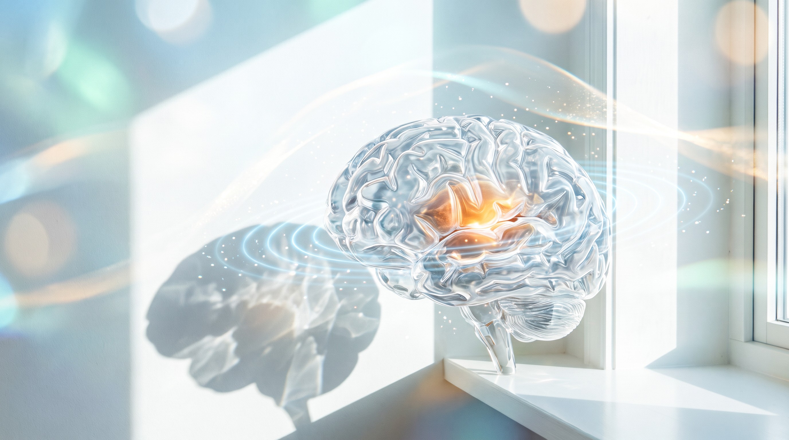
NeuroQuant is an FDA-cleared software tool that performs automated volumetric analysis of brain MRI. It takes a specific kind of three-dimensional scan, a high-resolution T1-weighted sequence, and it segments the brain into individual structures. It then calculates the volume of each structure and compares that to a normative database of people matched by age, sex, and head size.
The result is a report showing you percentiles. Your hippocampus may sit at the 12th percentile. Your amygdala may be at the 80th. Your thalamus may be at the 40th. Your ventricles, the fluid spaces in the brain, may be bigger than expected.
Other similar tools exist. The principle is the important thing. We are moving from a verbal impression to a measurement.
Here are some of the structures I often look at, and why.
Hippocampus. Central to memory formation and to placing experiences in context. It is especially vulnerable to chronic stress, poor oxygenation, and Alzheimer disease.
Amygdala. Involved in threat detection and emotion. Volume changes have been studied in trauma, mood disorders, and neurodegeneration.
Thalamus. The great sensory relay station of the brain.
Forebrain parenchyma. The overall volume of cortical and subcortical gray matter and white matter.
Cerebellum. Important in balance, coordination, timing, and emotional regulation.
Lateral ventricles and the inferior lateral ventricles. When brain tissue shrinks, fluid spaces can expand.
Basal ganglia, including the caudate and the putamen. Important in movement, reward, and motivation.
What This Can Tell Us
Volumetric analysis is most firmly established in memory disorders. Hippocampal atrophy is a recognized imaging marker in Alzheimer disease, and the FDA clearance of such tools reflects their role in quantifying brain volumes for clinical interpretation. Newer amyloid-targeting medications have added further interest in precise imaging.
Measurement also offers a way to detect patterns. Different neurodegenerative conditions can affect different regions, and a volumetric report can draw attention to an asymmetry or a pattern that would be easy to miss.
Traumatic brain injury is another important area. A growing body of research reports volumetric changes after brain injury, sometimes with atrophy that develops over time and sometimes with asymmetries. I have to be careful, though. An atrophy pattern cannot by itself prove that an injury caused it, particularly in a legal setting. Yet for a person who has been told that their scan is normal and that their symptoms must be psychological, an objective finding can feel like being believed for the first time.
Then there is toxic burden. In the field of biotoxin illness, some clinicians have reported characteristic volumetric patterns in patients exposed to water-damaged buildings, such as changes in the caudate, the pallidum, and the forebrain. This remains an area of working hypothesis, and these findings have not been established broadly. I treat them as a clue and never as a verdict. Still, I find it helpful to have an objective measure when someone has foggy thinking and a moldy home.
What This Cannot Tell Us
I would be doing you a disservice if I presented this as a crystal ball. Here are its limits.
A volume is not a function. Two people can have the same hippocampal volume and very different memories. One person may have enormous cognitive reserve built through a lifetime of education, music, and rich relationships.
A number does not name a disease. NeuroQuant does not diagnose Alzheimer disease, mold illness, or a concussion. A low percentile tells us where to look. It does not tell us why.
Scanner differences matter. Results can vary somewhat across different MRI machines and software versions. If we are tracking change over time, the same scanner and protocol are ideal.
Low percentiles do not always mean that something is wrong. In any large population, somebody has to be at the fifth percentile. Normal variation exists, and interpretation must fit your clinical story.
An enlarged structure is not always good. A swollen region may reflect inflammation rather than health. Conversely, a large region may simply be how you are built.
Results can also be frightening if they are delivered badly. A person receiving a report with red flags should have it explained thoroughly by someone who will also give them a plan.
I never treat a scan. I treat a person, and the scan is one voice in the conversation.
Function Plus Structure
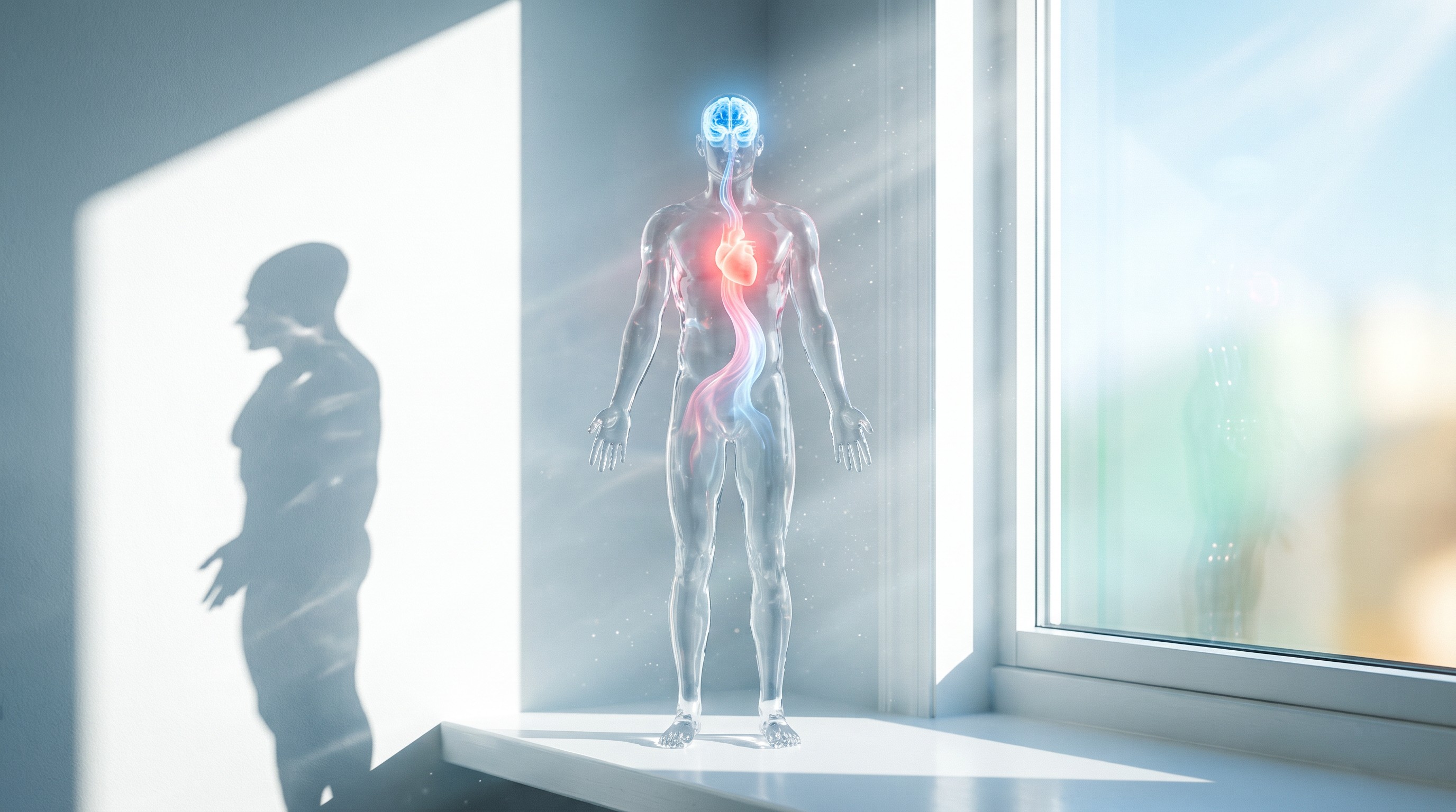
This is where the real power emerges. A volumetric MRI shows me structure. A comprehensive functional neurological exam shows me function. Together, they create a three-dimensional picture.
Let me give you an example of how the logic works. If a report shows a reduced right cerebellar volume, I want to look at the balance, the eye movements, and the coordination on that side. If the exam matches the scan, my confidence rises. If they do not match, I ask why. If the hippocampus is at a low percentile and memory testing is excellent, I look for what has been compensating so well. If the hippocampus is normal but the patient reports trouble, I look at attention, sleep, and metabolism.
A complete picture also includes biochemistry: fasting insulin, homocysteine, B12 with methylmalonic acid, thyroid panel, ferritin, vitamin D, omega-3 index, inflammatory markers, and toxic burden. I may assess cognition with structured tools. I may ask about sleep apnea, which is a major and treatable threat to the hippocampus.
Imagine that you are restoring a historic house. Volumetrics tells you the current dimensions of the rooms. The functional exam tells you whether the lights turn on. The biochemistry tells you whether the wiring and plumbing can support what you want to do.
The Most Hopeful Fact in Neuroscience
For most of the twentieth century, the common belief was that the adult brain was fixed, and that you could only lose ground. That belief turned out to be wrong.
One landmark line of research looked at London taxi drivers, who memorize thousands of streets. Parts of their hippocampi were larger than those of comparison groups, and the size correlated with how long they had been driving. Their brains appeared to have physically adapted to the job.
A randomized trial among older adults found that a year of moderate aerobic exercise increased the volume of the anterior hippocampus by about two percent, while those in a stretching group lost volume. Think about that. A year of brisk walking appeared to reverse a measure of age-related hippocampal shrinkage.
Other research has associated cognitive training, meditation, and treatment of sleep apnea with structural changes in the brain. The extent of adult human neurogenesis is still under active scientific debate. Yet there is no serious debate that the adult brain remains plastic.
So a volumetric scan is not a sentence. It is a baseline. That is the single most important way to think about it.
When we build a plan to reduce neuroinflammation, increase neurogenesis, and rebalance neurotransmitters, it can be powerful to have a starting point. I cannot promise that a volume will grow. I can tell you that the biology gives us every reason to work on it.
What Shrinks a Hippocampus, and What Feeds It
The hippocampus may be the most sensitive structure in the human brain. It sits at the intersection of memory, stress, oxygen, and metabolism, which makes it an honest reporter of how a life is being lived.
It is packed with glucocorticoid receptors, so chronic cortisol exposure can act upon it directly. People with Cushing syndrome, whose bodies make far too much cortisol, have shown reduced hippocampal volume, with some recovery after treatment. That finding alone tells you something profound. A hormone can change the size of a memory structure, and removing the excess can begin to reverse it.
It is exquisitely sensitive to oxygen, which is why untreated sleep apnea is such a serious concern. Studies have reported brain changes in people with apnea, and some have shown improvement following effective treatment.
It is sensitive to blood sugar. Higher blood sugar has been associated with smaller hippocampal volumes, even in people without diabetes. It is sensitive to high homocysteine and to inadequate B vitamins. It is also sensitive to alcohol, to chronic inflammation, to untreated hearing loss, and to social isolation.
Now consider what feeds it. Aerobic exercise raises brain-derived neurotrophic factor. Deep sleep supports memory consolidation. Novelty, including travel, a new language, or a new musical instrument, gives it something worth encoding. Close relationships appear to protect it. Omega-3 fats and a diet rich in colorful plants are associated with better brain aging in observational research.
Each item on that first list is a measurable thing, and each item on the second is something you can do this week. That is what I mean when I say that a brain scan is a beginning.
Hearing deserves particular mention. A large randomized trial reported that hearing aids slowed cognitive decline among a subset of older adults at higher risk. If your family has been asking you to turn the television down, your brain may benefit from that appointment.
When I review a volumetric report with a patient, I go through these lists together. We do not stare at a percentile and feel afraid. We ask what is costing this structure, and what we can give it.
Why Baselines Matter Most Before You Need Them
Bone density is measured before a fracture. Cholesterol is measured before a heart attack. The most useful moment for a brain baseline may be while you still feel fine. Changes in brain structure can begin well before symptoms, and a comparison between two scans tells far more than a single snapshot ever can.
Who Might Benefit From One
Not everyone needs a volumetric MRI. Here are the situations where I am most likely to consider one.
A person with concerns about memory or word-finding, particularly in midlife
A strong family history of dementia, in someone who wishes to work on prevention early
A concussion or brain injury with lasting symptoms, or several head injuries over a lifetime
Former contact sport athletes and military veterans who were exposed to repeated impacts or blasts
Cognitive symptoms after exposure to a water-damaged building
Unexplained neurological or cognitive change that has not been resolved by routine evaluation
People with sleep apnea, diabetes, or hypertension, who are at elevated risk and want a motivating baseline
An MRI is not without its practical burdens. It needs an order from an authorized clinician, it may not be covered by insurance, and it is not safe for everyone. Certain implants and devices may be incompatible. Claustrophobia can be a real obstacle. Children and pregnant patients need a separate conversation, and routine volumetric imaging is rarely called for in those groups.
A red flag remains a red flag. Sudden neurological changes need urgent medical care, not an elective volumetric study.
How to Ask for It
If you are getting an MRI, or planning one, it helps to ask ahead of time. NeuroQuant requires a three-dimensional volumetric T1 sequence, which a standard protocol may not include. Asking the imaging center whether they can perform this sequence, and whether they can run the volumetric analysis, can save you from needing to repeat the scan.
Ask to keep a copy of your images on disc or through a digital portal. In many cases, a prior scan that included the proper sequence can be processed retrospectively. A scan from five years ago, if it was captured with the right settings, can become an invaluable point of comparison.
Then make sure that someone can actually interpret the report in the context of your whole life. A report with percentiles and no plan may only increase your worry.
Children and the Developing Brain
I rarely need an MRI to understand a child. Most of what I need is visible in how they move, how their eyes track, how they feel, and what their reflexes are doing. Children are also more likely to need sedation for a scan, and I do not take that lightly.
However, I find the broader lesson important. The brain develops in a measurable sequence. Regions supporting movement and sensation mature early, while the prefrontal cortex does not fully mature until a person is in their mid-twenties. Sports concussions in adolescents happen during a critical phase of this construction. So I urge families to take head injuries seriously, even when the routine scan is clear.
During pregnancy, I focus on reducing exposures and building nutritional reserves. The structures that appear in a report at the age of sixty began to form in the womb.
Our brain is the master system, at any age.
Numbers Can Be Healing
I have seen the look on a face when someone sees their own data. For people who have spent years hearing that their scan was normal and that their problem must be stress, a clear finding can bring tears of relief. Someone has finally looked. Their experience has a shape.
I have also seen the opposite. A person terrified by a few memory lapses sees that their hippocampus sits at a healthy percentile and they exhale for the first time in a year. Their worry falls, and their memory often improves because of it.
Either way, the data serves the human being. It gives direction. It replaces fear with a map.
We tend to measure what we value. We measure our cholesterol, our bone density, our retirement account. We rarely measure the organ that holds the whole of who we are.
Your brain deserves a number and a plan.
Your baseline can be known. Your reserve can be built. Your future can be mapped with care.
Schedule Your Comprehensive Functional Neurological Exam Today
Book your Comprehensive Functional Medicine Eval/Neurological Exam today to discover what the contributing root causes are that are underlying your health concerns.
Begin at drleiladoolittle.com. I see patients by telehealth and in person in Dallas, Texas and Compton, Arkansas.

Comments